Technology
For combined orthodontic and surgical treatment to lead to the best possible dream smile with the greatest possible safety, certain technological foundations are necessary. In this respect, the Eltz Institut is a leader across Europe.
“Planning makes perfect”, as the saying goes. And it is no different with combined orthodontic and surgical treatment. That is why you need to get a good overview of the initial situation before straightening teeth and jaws – and then be able to translate this planning into reality:
- 3D cone beam CT (CBCT)
- Digital scan
- Digital panoramic X-ray
- Digital cephalometric X-ray
- Digital extraoral and intraoral facial photos
- 3D printing
- Special high-tech software for combined orthodontic and surgical treatment
3D cone beam CT scanner
With the help of a 3D cone beam CT scanner, the final position of your tooth roots in the bone can be planned three-dimensionally for the first time, which means damage to the tissues supporting your teeth (periodontium) can be avoided.
Incidentally, this device also makes us one of the first orthodontic centres in Europe able to simulate operations in advance and show the resulting changes to the face. Particularly in combination with oral and maxillofacial surgery, this allows us to achieve a depth of diagnostic planning that is unique.
Digital scan
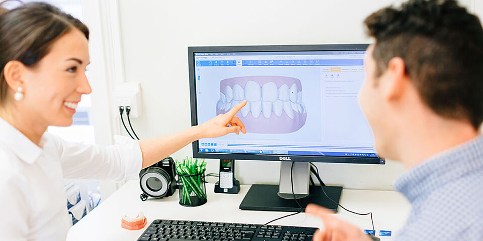
A digital scan gives us virtual models of the teeth and an accurate picture of the actual bite. In the past, this required taking impressions with cold metal trays in the mouth and sticky alginate or silicone material. Patients found this very unpleasant.
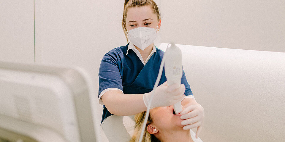
With our digital scan, everything is now much more comfortable: we move the scanner wand along the rows of teeth and the built-in laser transfers the data to the computer. Nothing escapes the sensitive scanner head: working at top speed and in high resolution, the scan captures even irregular surfaces with great precision.
Digital panoramic X-ray
On the one hand, the panoramic X-ray helps us assess your teeth and tooth roots, your jaw joints, your sinuses and the general condition of your bones, as well as how your teeth are positioned in the jaw.
On the other hand, the X-ray also gives us clues to possible difficulties in your treatment: Is there perhaps a tooth still hidden in the jaw that has not yet erupted (i.e. become visible) – and therefore has to be removed before other teeth are moved into that space? Are there abscesses, cysts or tumours (rare, of course, but this has to be checked too)? Are there teeth that we must not move under any circumstances, such as implants, bridges or baby teeth? Do we see any risks to your tooth roots, which could die off if treated?
This information already provides another good basis for planning your treatment. But we need considerably more information – such as the cephalometric X-ray.
Digital cephalometric X-ray
When your teeth are moved through the bone into a new position with orthodontic treatment, we need to know in advance how your teeth are anchored in the bone. This is because your teeth may only be moved to where there is a healthy periodontium and, above all, sufficient bone. After your braces, too, your teeth should once again be securely surrounded by bone, in order to prevent receding gums and keep your teeth healthy for the long term.
For this, we use a second X-ray, the so-called lateral cephalometric X-ray (FRS).
Using this two-dimensional side-view X-ray, we assess how your jaws are positioned in relation to each other and how your front teeth sit in the bone when viewed from the side. This lets us estimate how thick the layer of bone is around your incisors, for example. We also deduce in which direction your teeth may be moved – or, above all, must not be moved. We absolutely must avoid moving your teeth outside the safe area of the jawbone. Otherwise your gums could recede, your periodontium could be damaged and, in the worst case, you could even lose some of your teeth.
Digital intraoral and extraoral facial photos
Photographs of your face (extraoral photos) help us plan your treatment. While the photo from the side provides information about your profile, your lip closure, your lip position and your facial type, the frontal photo is used to examine disproportions and shifts in symmetry. In other words: we look for the actual reason why your smile is not yet Hollywood-ready. To make teeth end at the same height, for example, we can move some teeth upwards. Or others downwards. These are decisions we have to make.
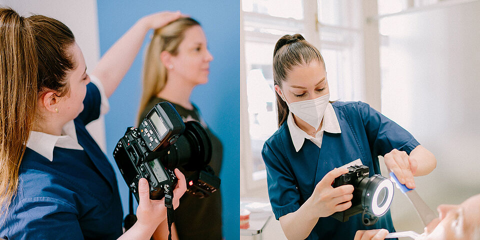
Photos of the inside of the mouth (intraoral photos), on the other hand, document the initial situation and will later show you the progress of your treatment.
Don’t leave your dental health to chance – take advantage of the revolutionary technologies at the Eltz Institut
Your consultation with us is free of charge and without obligation. We look forward to your online booking!